The brief
Connect sample preparation to the importance of observation depth. After one real laboratory shot, spend eighteen seconds traveling through an independent original field of cell-like forms, with different layers coming into visual focus. Preserve the distinction between an acquired image, an illustrative teaching view and a complete interpretation.
Audience: training teams, technical reviewers and process owners creating a concise microscopy focus and sample context film.
The visual approach
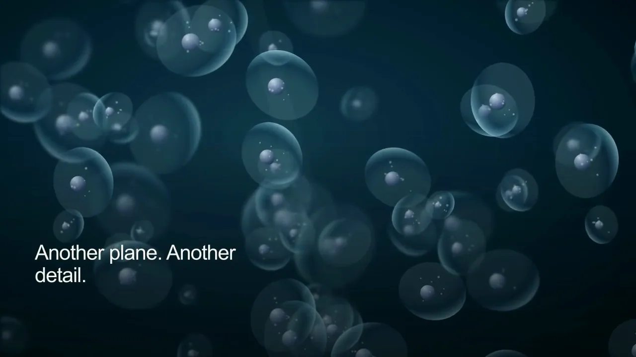
00–06s: A researcher prepares a specimen at the microscope. 06–12s: An original blue field contains distinct foreground and distant cell-like layers. 12–18s: Fictional nearby forms sharpen while distant forms soften through an independent focus-inspired transition. 18–24s: The motion passes through the layered fictional field, with no measured result or named organism.
The airy composition uses a 76 BPM rhythm selected for this storyboard. On-screen words stay short so the microscopy focus and sample context imagery carries the sequence.
Review and editable scope
A real production brief would keep the microscopy focus and sample context subject, source attribution, exact narration and caption wording available for review. A subject specialist must approve the factual explanation before using it in a real lesson.
The studio production plan keeps the four shot selections, crop, title, narration, captions and sound design separate. These notes document how this authored film was made. Use the Martius feature documentation for the product editing and review workflow.
Sources and interpretation
The specimen preparation is licensed laboratory stock. The cell-like forms, internal details, depth positions and moving focus are original fictional motion design, not an acquired microscopy image, named specimen, optical simulation, validated focusing method or instrument operating procedure.
The linked NIH / NIBIB: optical imaging and microscopy resource supports the conceptual topic. Stock subjects are illustrative and are not Martius customers, endorsements or evidence of a named facility’s practices. Original illustration is marked as conceptual wherever its appearance could be confused with observed data.
Scene-by-scene transcript
00–06s — A researcher prepares a specimen at the microscope. On-screen text: Start before the image.. Narration: The sample and its record travel into microscopy. Original stereo score.
06–12s — An original blue field contains distinct foreground and distant cell-like layers. On-screen text: A view selects a depth.. Narration: Focus changes which structures appear sharp in the view. Original stereo score.
12–18s — Fictional nearby forms sharpen while distant forms soften through an independent focus-inspired transition. On-screen text: Another plane. Another detail.. Narration: Different depths reveal a different view of the sample. Original stereo score.
18–24s — The motion passes through the layered fictional field, with no measured result or named organism. On-screen text: Keep the context with the view.. Narration: Interpretation needs the sample context and acquisition record. Original stereo score.
Words, sound and scene notes
- 00:00
- A researcher prepares a specimen at the microscope. On-screen text: Start before the image.. Narration: The sample and its record travel into microscopy. Original stereo score.
- 00:06
- An original blue field contains distinct foreground and distant cell-like layers. On-screen text: A view selects a depth.. Narration: Focus changes which structures appear sharp in the view. Original stereo score.
- 00:12
- Fictional nearby forms sharpen while distant forms soften through an independent focus-inspired transition. On-screen text: Another plane. Another detail.. Narration: Different depths reveal a different view of the sample. Original stereo score.
- 00:18
- The motion passes through the layered fictional field, with no measured result or named organism. On-screen text: Keep the context with the view.. Narration: Interpretation needs the sample context and acquisition record. Original stereo score.