The brief
Explain the echo principle through a clearly schematic transducer and wave motion, without showing a diagnostic result.
Audience: educators, technical communicators and subject specialists creating a concise ultrasound and medical imaging film.
The visual approach
00–06s: A schematic transducer releases expanding curved wave fronts. 06–12s: A distinct warm marker travels back through the layered scene. 12–18s: Outgoing waves and returning markers remain visually separate. 18–24s: The full layered conceptual view closes without depicting a clinical scan.
The science composition uses a 80 BPM rhythm selected for this storyboard. On-screen words stay short so the ultrasound and medical imaging imagery carries the sequence.
Review and editable scope
A real production brief would keep the ultrasound and medical imaging subject, source attribution, exact narration and caption wording available for review. A subject specialist must approve the factual explanation before using it in a real lesson.
The studio production plan keeps the four shot selections, crop, title, narration, captions and sound design separate. These notes document how this authored film was made. Use the Martius feature documentation for the product editing and review workflow.
Sources and interpretation
Original waves, tissue bands and echo markers are conceptual and not anatomically scaled, physically timed, patient-specific or intended for diagnosis.
The linked NIH / NIBIB: ultrasound resource supports the conceptual topic. Stock subjects are illustrative and are not Martius customers, endorsements or evidence of a named facility’s practices. Original illustration is marked as conceptual wherever its appearance could be confused with observed data.
Scene-by-scene transcript
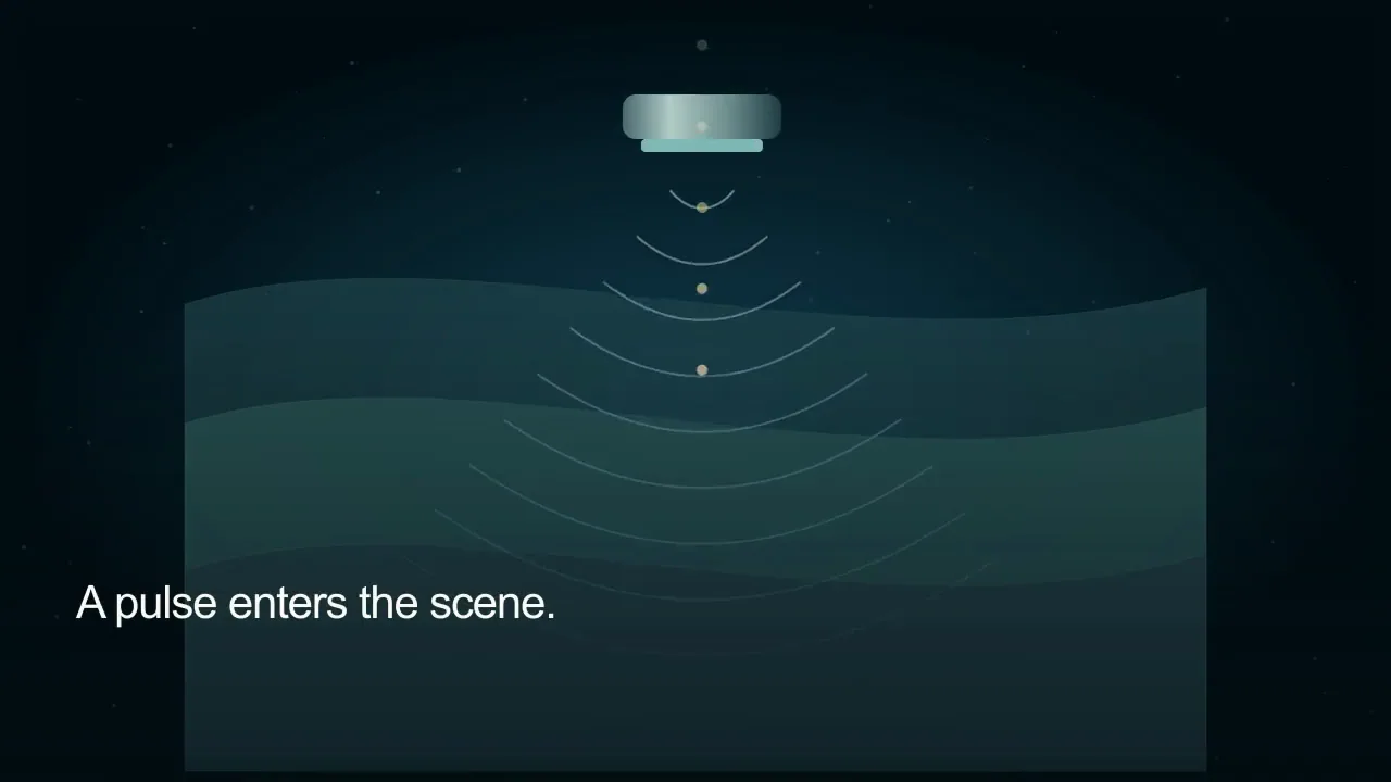
00–06s — A schematic transducer releases expanding curved wave fronts. On-screen text: A pulse enters the scene.. Narration: A transducer sends high-frequency sound into tissue. Original stereo score.
06–12s — A distinct warm marker travels back through the layered scene. On-screen text: Boundaries return an echo.. Narration: Tissue boundaries reflect some of that sound. Original stereo score.
12–18s — Outgoing waves and returning markers remain visually separate. On-screen text: Timing tells a distance.. Narration: Echo timing helps estimate the boundary’s distance. Original stereo score.
18–24s — The full layered conceptual view closes without depicting a clinical scan. On-screen text: Echoes help build a view.. Narration: Returned signals help construct an ultrasound image. Original stereo score.
Words, sound and scene notes
- 00:00
- A schematic transducer releases expanding curved wave fronts. On-screen text: A pulse enters the scene.. Narration: A transducer sends high-frequency sound into tissue. Original stereo score.
- 00:06
- A distinct warm marker travels back through the layered scene. On-screen text: Boundaries return an echo.. Narration: Tissue boundaries reflect some of that sound. Original stereo score.
- 00:12
- Outgoing waves and returning markers remain visually separate. On-screen text: Timing tells a distance.. Narration: Echo timing helps estimate the boundary’s distance. Original stereo score.
- 00:18
- The full layered conceptual view closes without depicting a clinical scan. On-screen text: Echoes help build a view.. Narration: Returned signals help construct an ultrasound image. Original stereo score.